Technology
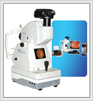 |
Fundus Camera/ Retinal Camera A fundus camera or retinal camera is a specialized low power microscope with an attached camera designed to photograph the interior surface of the eye, including the retina, optic disc, macula, and posterior pole (i.e. the fundus). Fundus/ retinal examination plays an important role in the diagnosis and management of several medical conditions like diabetes, hypertension, and raised intracranial pressure among others. |
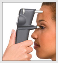 |
Tonometer Tonometer measures the Intraocular Pressure (pressure inside the eye). Traditionally, this was done by using a local anaesthetic, resting a weight on the cornea and measuring how much the cornea could be depressed. This works on the principle that the force required to flatten a given area of the cornea is proportional to the pressure inside the eye. This is one of the most important tools in an eye examination used for the early detection of glaucoma. The non contact tonometers we use do not touch the cornea and do not require the use of a local anaesthetic. It gives the measurement using a puff of air, and only filtered air touches the cornea. |
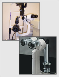 |
Slit Lamp A slit lamp is an examination device that consists of a moveable light source and binocular microscope for viewing the external eye structures such as the cornea, conjunctiva, sclera and lids. It is an essential tool in contact lens fitting and management and for detecting cataracts then documenting these images digitally. It is important for us to monitor changes over time, especially with pathologies and cataracts, and by documenting these digital images we can compare and quantify these changes. |
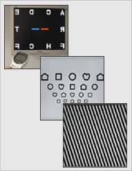 |
Computerized Letter Charts Computerized Letter Charts allow accurate refractions by randomly generating either numbers, letters, symbols or pictures and by changing the contrast levels. The eye detects objects by responding to the differing levels of illumination at the edges of visual targets. Contrast acuity or sensitivity, therefore, has to do with the ability of the eye to resolve a given target against the background illumination. The closer the levels of target illumination and background illumination, the more difficult it becomes to resolve and hence identify the target. A reduction of contrast acuity occurs in many eye disorders, especially corneal diseases, cataracts, and retinal degenerative disorders. |
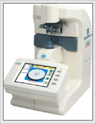 |
Computerized Spectacle Lens Analyzer This is used to check your new and/or previous spectacle lenses for errors and distortions, with prescription accuracy of 100th of a diopter. It also allows us to determine if the positioning of your lenses were done correctly. This is used to check your new and/or previous spectacle lenses for errors and distortions, with prescription accuracy of 100th of a diopter. It also allows us to determine if the positioning of your lenses were done correctly. |
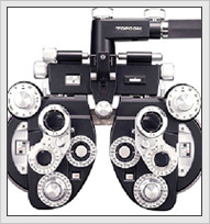 |
Phoropter This incorporates a whole battery of different types of lenses and prisms to accurately determine your refractive needs and check on your distance and reading prescription, focus ability, binocular vision and stereoscopic vision. |
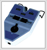 |
Specialized Pupil Distance Measure Meter This is essential for high prescriptions and progressive bifocal (multifocals) lens fitting. It allows us to measure very accurately your eye position on either side of your nose bridge which ensures that you have the best vision possible with progressive lenses.
|
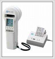 |
Corneal Surface Measurement A keratometer is primarily used to diagnose the presence of astigmatism and to determine the degree and treatment of astigmatism. Our Nidek KM-500 keratometer is a new generation medical instrument that can also measure the curvature and reflection of the anterior surface of the cornea. The measurement of cornea curvature is extremely important for the selection of contact lens base curve (B.C.) in the contact lens fitting procedures.
|
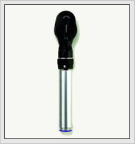 |
Ophthalmoscope An ophthalmoscope is used to examine the interior of the eye, including the lens, retina and optic nerve. The ophthalmoscope consists of a concave mirror and a battery-powered light (contained within the handle). The optometrist looks through a single monocular eyepiece into the patient's eye. The ophthalmoscope is equipped with a rotating disc of lenses to permit the eye be examined at different depths and magnifications. |